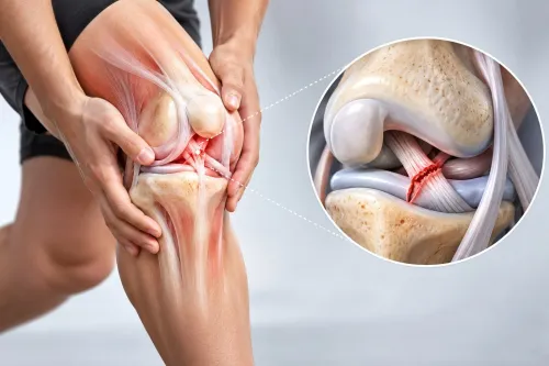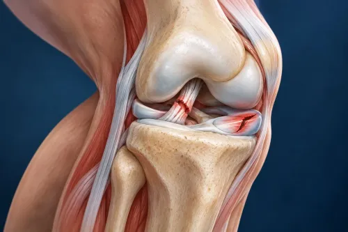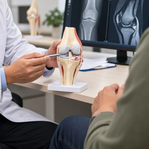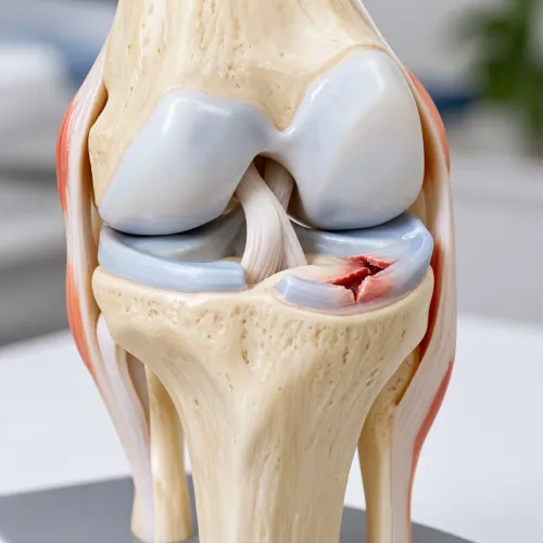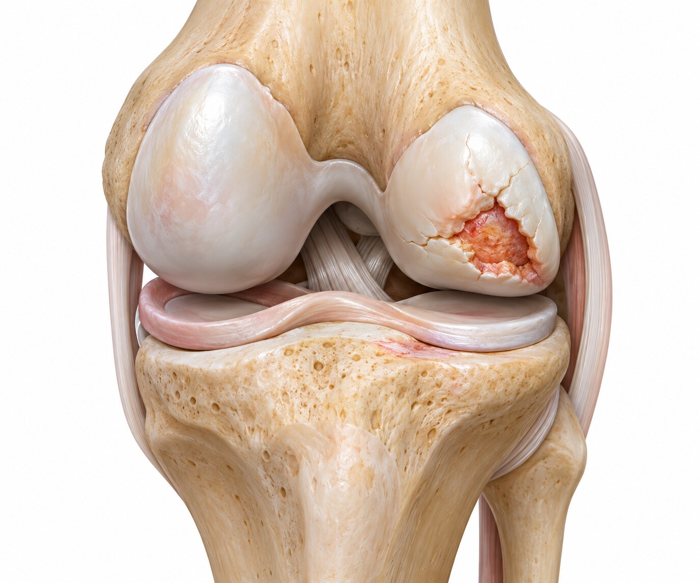
A small area of damaged knee cartilage can cause pain, swelling, catching, and restricted movement even when the rest of the joint remains relatively healthy. By the end, you will understand how doctors identify a true focal defect, match its size and location to a treatment, and judge whether repair offers a better option than rehabilitation or joint replacement.
Key takeaways
- Localized defects often hurt during specific movements or after repeated loading.
- MRI helps measure the lesion and identify associated meniscus, ligament, or alignment problems.
- Procedure choice depends on defect size, depth, location, symptoms, and patient factors.
- Rehabilitation takes months and requires gradual progression before impact activities.
What localized cartilage damage feels like
A focal cartilage defect usually produces pain in one repeatable spot, triggered by a particular load or movement, rather than constant discomfort throughout the knee. Localized cartilage damage can cause pain with stairs or squatting, swelling after activity, catching, grinding, or difficulty fully bending and straightening the joint.
These knee cartilage symptoms become more suspicious when the knee feels reasonably normal during rest.
| Pattern | Typical clues | What it suggests |
|---|---|---|
| Focal cartilage defect | Localized pain, activity-related swelling, catching, or pain during a specific movement | Damage in one cartilage area, often after injury or repeated overload |
| Broader cartilage loss | Stiffness after rest, pain across the joint, reduced walking tolerance, and symptoms in several activities | Osteoarthritis affecting a compartment or multiple knee structures |
| Meniscus problem | Joint-line tenderness, twisting pain, catching, or locking | Meniscal tear rather than cartilage damage alone |
| Ligament problem | Giving way after a pivot, sudden direction change, or landing | Instability, especially after a ligament injury |
| Bone or inflammatory problem | Deep constant pain, night pain, marked warmth, prolonged stiffness, or fever | Bone injury, infection, or inflammatory arthritis requiring separate assessment |
A scan showing a defect does not prove it causes the pain. Widespread arthritis, a meniscal tear, poor kneecap tracking, malalignment, ligament instability, or damaged bone can create similar symptoms or prevent a focal treatment from working. Examination and weight-bearing X-rays help distinguish an isolated lesion from a broader knee problem.
How doctors confirm the defect and its cause
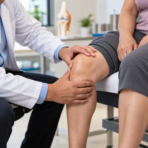
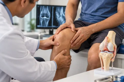
A knee physical examination helps determine whether the defect matches your pain. Your clinician checks tenderness, swelling, range of motion, creaking, patellar tracking, ligament stability, alignment, and pain during squatting or resisted movement. They also compare the pattern with meniscus tear symptoms, such as joint-line tenderness, catching, or pain during twisting.
| Test | What it shows | What it cannot prove |
|---|---|---|
| Standing weight-bearing X-ray | Alignment, joint-space narrowing, bone spurs, fractures, and arthritis under load | Cartilage itself; a normal-looking joint space does not exclude a focal defect |
| Knee cartilage MRI | Cartilage thickness, surface fissures, full-thickness loss, bone marrow changes, menisci, ligaments, and swelling | That the visible lesion is the pain source |
| Diagnostic assessment | Whether symptoms, examination findings, and imaging point to one area | A scan alone cannot separate an incidental defect from the true cause |
Doctors build the diagnosis by matching all three sources. Pain that occurs when the damaged area is loaded, local tenderness, recurrent swelling, and restricted motion strengthen the link; widespread pain, marked arthritis, instability, malalignment, or inflammatory signs point elsewhere.
A local anaesthetic injection into the joint can provide additional evidence when the pain source remains uncertain. Surgeons also review whether symptoms improve with activity modification and strengthening before recommending cartilage repair.
Which cartilage procedure matches the lesion
A small, contained full-thickness defect in a healthy knee may suit cartilage restoration; a large defect, damaged bone, or widespread arthritis changes the decision. Lesion depth matters because surface-only wear does not need the same operation as cartilage loss reaching subchondral bone.
| Procedure | Best match | Main limitation |
|---|---|---|
| Microfracture knee surgery | Small, contained defects, especially in younger or lower-demand patients | Forms fibrocartilage rather than native hyaline cartilage and is less reliable for large lesions |
| Osteochondral autograft transfer, called OATS or mosaicplasty | Small to medium, accessible defects needing mature cartilage and bone plugs | Donor tissue is limited and the harvest site can become painful |
| Osteochondral allograft transplantation | Larger defects or substantial bone involvement | Requires a suitable donor graft and involves more complex surgery |
Location also matters. A defect on a weight-bearing surface faces greater load than one in a less-loaded area, while patellar or trochlear lesions require careful assessment of tracking. Alignment, ligament stability, and meniscus health determine whether the repair has a fair mechanical environment.
Corrective osteotomy, ligament reconstruction, meniscal repair, or meniscal transplantation may need to accompany cartilage treatment.
Age, occupation, and sport demands influence durability and rehabilitation expectations, but they do not decide eligibility alone. Replacement surgery becomes more appropriate when arthritis affects multiple compartments, the knee is severely malaligned or unstable, or meniscal and bone loss cannot support a focal repair. A visible MRI lesion alone is not enough.
What happens during treatment and rehabilitation
Through two or three small knee arthroscopy portals, the surgeon inserts a camera and instruments to inspect the defect, remove loose fragments, and treat its surface. Arthroscopic chondroplasty and cartilage debridement trim unstable flaps and smooth rough edges; they can reduce catching, but they do not recreate normal cartilage.
| Procedure | Weight-bearing and physiotherapy | Typical return to sport |
|---|---|---|
| Chondroplasty or debridement | Walking often progresses quickly, unless another procedure was performed. Physiotherapy restores motion, swelling control, and strength. | Lower-impact activity may return within weeks; pivoting sport depends on pain, strength, and the rest of the knee. |
| Microfracture | Crutches and protected weight bearing commonly last several weeks while marrow clot forms over the defect. Early physiotherapy protects the repair while maintaining motion. | Running and pivoting often wait several months, with progression based on strength and symptoms. |
| Osteochondral graft | Weight-bearing is restricted while the plug or donor graft heals. Physiotherapy increases motion and loading according to graft location and fixation. | Sport commonly takes many months; contact and pivoting begin only after objective strength and stability testing. |
| Autologous chondrocyte implantation or MACI | This is staged: cells are harvested during one arthroscopy, expanded, then implanted later. Autologous chondrocyte implantation recovery involves prolonged protected loading and structured rehabilitation. | Return to pivoting sport often takes 9–12 months or longer, not weeks. |
The lesion’s location, graft fixation, and any ligament, meniscus, or alignment operation set the actual restrictions. A successful repair still needs gradual loading; rushing sport can damage the healing surface.
How to decide whether cartilage repair is worthwhile
Cartilage repair is worthwhile when a symptomatic, localized full-thickness defect sits in an otherwise preserved, stable knee. It can reduce pain, swelling, and activity limits, but it cannot promise normal cartilage, permanent relief, or protection from future arthritis.
Nonoperative care should come first when symptoms are manageable or the knee has diffuse osteoarthritis. Physiotherapy, strength training, weight management, activity changes, and appropriate pain relief may control symptoms without surgical risk. A focal repair is unlikely to work if untreated malalignment, ligament instability, meniscus loss, or inflammatory disease continues loading the defect.
| Option | Realistic benefit | When it comes first |
|---|---|---|
| Nonoperative care | Better strength, movement, and symptom control | Mild symptoms or widespread arthritis |
| Cartilage repair or restoration | Improved symptoms and joint function; tissue may not match native cartilage | Focal defect with preserved surrounding joint |
| Partial knee replacement | Reliable pain relief for arthritis confined to one compartment | Compartment-level cartilage loss, not an isolated defect |
The distinction between cartilage repair versus restoration matters. Microfracture produces mainly fibrocartilage versus hyaline cartilage, while graft procedures can transfer hyaline cartilage but have donor-site limits. Chondroplasty smooths unstable flaps; it does not regenerate cartilage.
Discuss knee cartilage surgery risks, including persistent pain, stiffness, infection, blood clots, graft failure, worsening arthritis, and revision surgery. Ask Dr Ronak Kothari how lesion size, alignment, meniscus health, and your return-to-sport goals affect the choice.
- What happens if I continue nonoperative treatment?
- Is partial knee replacement versus cartilage repair more appropriate?
- How long could the repair last, and what restrictions follow?
Related services
Knee Preservation Procedures Dr. Ronak Kothari, an experienced Orthopaedic Surgeon, specializes in Knee Preservation Procedures aimed at maintaining the natural knee joint for as... View service → |
Knee Arthroscopy & Sports Medicine Knee Arthroscopy & Sports Medicine Dr. Ronak Kothari, a skilled Orthopaedic Surgeon, specializes in Knee Arthroscopy and Sports Medicine, offering advanced, minimally invasive... View service → |
Frequently asked questions
What does localized cartilage damage in the knee feel like?
It often causes pain in one repeatable spot during stairs, squatting, running, or another specific load. Swelling after activity, catching, grinding, and reduced bending or straightening can also occur.
How do doctors confirm a cartilage defect and its cause?
Doctors use your symptom pattern, physical examination, weight-bearing X-rays, and MRI. These tests help identify the defect and check for meniscus injury, ligament damage, bone changes, or leg-alignment problems.
Which cartilage repair procedure is suitable for a knee lesion?
The choice depends on the defect’s size, depth, location, surrounding bone, activity goals, and overall knee condition. Options include marrow stimulation, osteochondral grafting, and autologous chondrocyte implantation.
What happens during recovery after cartilage repair surgery?
Recovery usually involves protected weight-bearing, early range-of-motion exercises, physiotherapy, strength training, and a gradual return to impact activity. The timeline depends on the procedure and lesion.
When is cartilage repair surgery worth considering?
It is worth discussing when a confirmed focal defect causes persistent symptoms despite appropriate non-surgical treatment, and the rest of the knee is suitable for repair.